MRI safety and hearing implants
Every patient with an implant, whether a hearing implant or hip prosthesis, receives a corresponding implant ID card, which he or she should show to the physician prior to medical examinations, if possible already when making an appointment. This gives doctors and patients enough time to obtain any additional information they may need about the implant in question.

Medical professionals are often in a difficult situation: not only are they responsible for the accuracy of their diagnoses and the success of the therapy, but they are also responsible for taking into account all the patient’ s history of treatments and illnesses, as well as his or her implants. Thus, the radiologist must know not only what type of imaging to use for optimal results, e.g. conventional X-ray, CT, MRI, ultrasound examination or other imaging, but also what interactions with the implant might occur in each case.
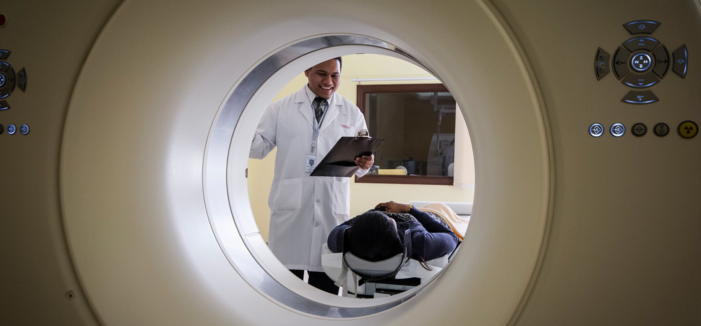
In general, X-ray or CT scans, as well as ultrasound examinations, are risk-free imaging procedures for users of hearing implants and the implants do not affect their quality. However, MRI examinations must be planned well in advance, since the mutual influence between hearing implant and MRI depends on the type of implant and must be taken into account.
The MRI safety of implants is becoming increasingly important. Prof. Ertl-Wagner explained at a symposium in April 2015 that every fifth German would need a so-called cross-sectional image every year, about 45% of which would be MRIs. In Austria, according to Prof. Dr. Wolf-Dieter Baumgartner, ENT University Clinic Vienna and president of the CIA, about 10% of all CI patients at AKH Vienna had to undergo an MRI in the last 19 years. He finds the MRI strength of hearing implants to be crucial, because, as he explains, a torn meniscus or ligament can quickly be contracted while skiing or on the soccer field, and “not every trauma surgeon at the ski resort knows what a vibrant soundbridge is.” Regular MRI exams are essential for patients with multiple sclerosis, NFII tumors, or similar comorbidities, emphasizes Prof. Martin Kompis, M.D., University Hospital of Bern. He is adding that it’s also a good idea to plan ahead “for those who don’t know if they’ll need an MRI in the next 60 or 70 years,” which ultimately includes every implant patient.
Interactions of MRI and hearing implant
MRI stands for “Magnet Resonance Imaging”, also called MRT “Magnet Resonance Tomography” or MR for short. In this process, strong alternating magnetic fields are generated, which exert a corresponding force on the magnetic parts of the implant. Now, there is not only a small magnet in the outer parts of hearing implant systems, which should be always taken off during the examinations, but also the implanted part of the system contains a magnet. Due to the interaction of this implanted magnet with the magnetic field of the MR device, the implant is subject to forces during imaging that could cause it to rotate or shift.
On the other hand, the magnet of the implant could lose its magnetic force. In addition, signals could be induced by the changing magnetic fields, creating unwanted, possibly unpleasant auditory sensations, and even heating of the human tissue in the area around the implant could be a consequence of the MRI. Still, the forces generated by the magnetic field are the main risks for the implant.
Moreover, not only is the implant with all its parts visible on the produced image, but it also casts a shadow directly in the implant area, which leads to distortions in the surrounding area. This means that in this area the informative value is impaired during MRI of the skull.
New generation of hearing implants
As a result of this, meaningful MR images of the implanted side of the skull are, with most hearing implants, not possible. However, MR images of users of MED-EL’s latest SYNCHRONY generation of cochlear-implants provide meaningful images, even of these ipsilateral structures in the skull when the magnet is removed for this purpose, as Dr. Omid Majdani from the Hannover Hearing Center recently demonstrated in a study.
DI Ewald Thurner, the director of MED-EL Vienna, explains: “With all our cochlear implants, as well as the Bonebridge and the new Vibrant Soundbridge VORP503, MR imaging up to 1.5 Tesla field strength is possible without safety risk, provided the manufacturer’s instructions are followed. The older Vibrant Soundbridge VORP502s are the only exception in this regard.” MRI compatibility is not the case with all manufacturers. Recently, not only is the overall frequency of MR diagnostics increasing, more and more MR machines with the field strengths up to 3 Tesla are being used.
“With 3 Tesla, it’s easier for radiologists to take good images,” explains Munich radiologist Prof. Dr. Birgit Ertl-Wagner. That’s why about 20 percent of all newly installed MR devices in Germany are 3T systems, she says. With some cochlear implants, it is also possible to make images with 3T if the magnet of the implant is removed beforehand. However, removing the magnet requires a minor surgical procedure and is associated with wound healing before the implant can be used again to its full extent. Prof. Baumgartner points out the cost relevance of magnet removal as well: “If you take into account various preliminary examinations, then a skin incision at a university hospital amounts to up to 5,000 euros.” From the point of view when MR diagnostics are necessary, an implant in which the tests can be performed without prior removal of the magnet shows many advantages. “MED-EL’s SYNCHRONY is the only CI in the world that is MR capable up to 3 Tesla without removing the magnet,” DI Thurner proudly explains.
But still, the described distortions occur in the area around the implant. So, if you need to see the tissue in that area precisely, the magnet has to be removed after all. DI Thurner comments, “This is also possible with SYNCHRONY.” In addition, it is possible to reduce the artifacts with certain settings of the MR devices, the so-called metal suppression sequences. In a study on artifacts in MR, Dr. Majdani found that the SYNCHRONY is the only CI in which MR examinations of the skull provide meaningful images even on the implanted side.
For all other images far from the skull, the magnet can remain in the implant. “There are no problems from the spine onwards,” explains Prof. Dr. Matthias Tisch, ENT surgeon at the German Armed Forces Hospital in Ulm. From there on, good quality images can be obtained without artifacts.
What to do?
When selecting the hearing implant, people should also pay attention to which current implants are MR-capable and to what extent. If MR is required, the radiologist should already be informed about the implant when making the appointment. Moreover, it should be clarified whether MR equipment with the required image sequences and field strengths is available in relation to the respective implant and whether the radiologist requires further information from the manufacturer of the implant. Such information can be obtained from the speech processor user manual, the implant identification card, the manufacturer’s website, or from the manufacturer directly. MED-EL also has an information sheet available which contains all necessary data. Waiting times for an MRI in Austria can be as long as 14 weeks, as Dr. Friedrich Vorbeck, chairman of the Bundesfachgruppe Radiologie, explains; in the private sector, it can be somewhat faster. Therefore, everything should be prepared so that the agreed appointment can then be kept.
Of course, alternative examination methods should sometimes be considered. Dr. Majdani refers to contrast media CTs for various questions, and Prof. Ertl-Wagner also mentions CT as a possible alternative. He immediately debunks the argument of higher radiation exposure with CT: “It is true that you have to pay attention to the dosage, but CT of ten years ago is not CT of today.” With careful adjustment, he said, the exposure with CT is about the same as with MRI. For a temporal bone CT, which images the particularly critical area around the hearing implant, the radiation is 0.11 millisieverts, which is equivalent to the radiation exposure on a flight from Munich to San Francisco.
If the decision to undergo MRI is already made, the user should remove the outer parts of the hearing systems for examination. MED-EL recommends a pressure bandage around the implant. The patient must lie on their back and not move their head during the examination.
MRI Guarantee for hearing implant users
From January 2021, MED-EL offers the global guarantee extension for the users of their implants. This guarantee covers possible damages to the implant which can arise as a result of MRI and which include implant damage, implant magnet dislocation, implant magnet demagnetization etc. Although these consequences are highly unlikely, the patients can receive the new implant or magnet free of charge from the manufacturer, in case these problems occur. Find all the details of the MED-EL MRI Guarantee here.
MED-EL is the only manufacturer to offer such a guarantee. The company has decades of positive experience with such examinations and points to the importance and urgency of such examinations: “Rapid access to MR examinations reduces the risk of misdiagnosis.”


